Retina
The retina is a layered structure with several layers of neurons interconnected by synapses. The only neurons that are directly sensitive to light are the photoreceptor cells. These are mainly of two types: the rods and cones. Rods function mainly in dim light and provide black-and-white vision, while cones support daytime vision and the perception of colour. A third, much rarer type of photoreceptor, the intrinsically photosensitive ganglion cell, is important for reflexive responses to bright daylight.
Retinal gene therapy
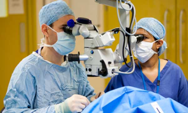
Gene therapy holds promise as a potential avenue to cure a wide range of retinal diseases. This involves using a non-infectious virus to shuttle a gene into a part of the retina. Recombinant adeno-associated virus (rAAV) vectors possess a number of features that render them ideally suited for retinal gene therapy, including a lack of pathogenicity, minimal immunogenicity, and the ability to transduce postmitotic cells in a stable and efficient manner. rAAV vectors are increasingly utilized for their ability to mediate efficient transduction of retinal pigment epithelium (RPE), photoreceptor cells and retinal ganglion cells. Each cell type can be specifically targeted by choosing the appropriate combination of AAV serotype, promoter, and intraocular injection site.
Retinitis pigmentosa
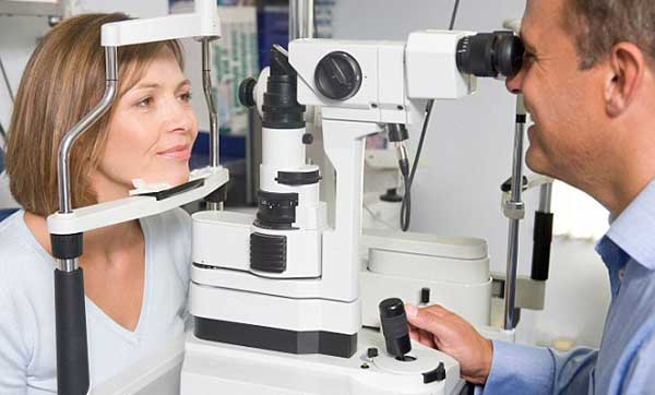
Retinitis pigmentosa (RP) is an inherited, degenerative eye disease that causes severe vision impairment due to the progressive degeneration of the rod photoreceptor cells in the retina. This form of retinal dystrophy manifests initial symptoms independent of age; thus, RP diagnosis occurs anywhere from early infancy to late adulthood. Patients in the early stages of RP first notice compromised peripheral and dim light vision due to the decline of the rod photoreceptors The progressive rod degeneration is later followed by abnormalities in the adjacent retinal pigment epithelium (RPE) and the deterioration of cone photoreceptor cells.
Evolution of the eye
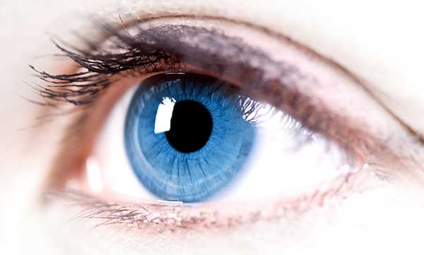
The evolution of the eye has been a subject of significant study, as a distinctive example of a analogous organ present in a wide variety of taxa. Complex, image-forming eyes evolved independently some 50 to 100 times. Complex eyes appear to have first evolved within a few million years, in the rapid burst of evolution known as the Cambrian explosion. There is no evidence of eyes before the Cambrian, but a wide range of diversity is evident in the Middle Cambrian Burgess shale, and the slightly older Emu Bay Shale. Eyes show a wide range of adaptations to meet the requirements of the organisms which bear them. Eyes vary in their visual acuity, the range of wavelengths they can detect, their sensitivity in low light levels, their ability to detect motion or resolve objects, and whether they can discriminate colours.
Retinal scan
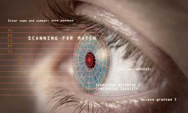
A retinal scan is a biometric technique that uses the unique patterns on a person's retina blood vessels. It is not to be confused with another ocular-based technology, iris recognition, commonly called an "iris scanner."
The human retina is a thin tissue composed of neural cells that is located in the posterior portion of the eye. Because of the complex structure of the capillaries that supply the retina with blood, each person's retina is unique. The network of blood vessels in the retina is not entirely genetically determined and thus even identical twins do not share a similar pattern.
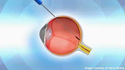
Visual prosthesis
A visual prosthesis, often referred to as a bionic eye, is an experimental visual device intended to restore functional vision in those suffering from partial or total blindness. In 1983 Joao Lobo Antunes, a Portuguese doctor, implanted a bionic eye in a person born blind. Many devices have been developed, usually modeled on the cochlear implant or bionic ear devices, a type of neural prosthesis.